
From detection to translation: How SPARC and ROSETTA are reshaping imaging-guided PCa management
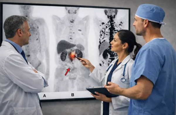
MRI and PSMA PET have changed the way we diagnose prostate cancer (PCa) over the past decade. Still, there is considerable variability in how imaging findings are interpreted, reported, and used in everyday clinical decisions. SPARC and ROSETTA are two initiatives that try to deal with these limitations from different angles.
Imaging has moved PCa diagnosis away from purely “general” approaches toward more selective strategies. Diagnostic pathways still differ between centres. SPARC was created to address this issue by focusing on how imaging is used in real life. Rather than promoting a single technique, SPARC looks at practical questions: who should undergo imaging, when biopsy is really needed, and how imaging results should influence management. In practical terms, SPARC helps us think more clearly about how to use imaging tools in a consistent way, aiming for better standardisation in PCa diagnosis and treatment.
Another key aspect of SPARC is its emphasis on how PSMA PET/CT should be reported. There was agreement on the value of structured reporting, including molecular PSMA expression levels, miTNM staging using the PROMISE criteria, and intraprostatic assessment tools such as the PRIMARY score. SPARC supported reporting quantitative parameters, such as PSMA tumour volume and standardised uptake values. There was agreement that SUVmax should be reported by region rather than as a single-lesion value, as this better reflects the extent and heterogeneity of disease.
At the same time, SPARC also made clear where uncertainty remains. The relationship between PSMA PET/CT findings and traditional risk groups in newly diagnosed metastatic PCa is still not fully understood. Likewise, there was no clear agreement that treatment decisions based only on PSMA PET/CT lead to better patient outcomes. SPARC also acknowledged that current evidence is not strong enough to define response or progression criteria based solely on PSMA PET/CT, underlining the need for further prospective studies.
ROSETTA, in contrast, focuses on interpretation. The Pelvic Rosetta Classification provides an interdisciplinary map of pelvic lymph node regions based on clear anatomical landmarks. Its main goal is simple: to ensure that urologists, radiologists, and nuclear medicine specialists are talking about the same areas in the same way. By translating imaging findings into well-defined pelvic regions, ROSETTA reduces ambiguity and makes imaging reports easier to apply in clinical practice. This is particularly useful in situations such as extended pelvic lymph node dissection, PSMA-radioguided surgery, and image-guided radiotherapy.
Overall, PCa management is moving away from a “one-size-fits-all” approach toward “haute-couture” personalised, image-guided decision-making. For the urologist, the key message is simple: SPARC and ROSETTA are not about adding complexity but about bringing more clarity to daily practice. The real value of advanced imaging lies not only in what it shows, but in how that information is used to guide patient care. As imaging continues to evolve, initiatives such as SPARC and ROSETTA help us to integrate innovation into clinical work, supporting better decisions along the prostate cancer pathway.
References
- The Pelvic Rosetta Classification Project: An Interdisciplinary Proposal for a Lymph Node Map of the Pelvis in Prostate Cancer.
Gernot Ortner, et al. Journal of Nuclear Medicine November 2025, jnumed.125.270667; DOI: 10.1016/j.eururo.2025.08.005 - SPARC: The Standardised Prostate-specific Membrane Antigen Positron Emission Tomography/Computed Tomography Analysis and Reporting Consensus: A Delphi Analysis
Ken Herrmann, et al. European Urology, Volume 89, Issue 3p260-274March 2026; DOI: 10.1016/j.eururo.2025.08.005